|
|
In this exhibit you learn about the ways we can look inside people's bodies to diagnose disease.
Body imaging is an important part of medicine. It is one of the ways that doctors can diagnose disease, that is, work out exactly what is wrong with a patient. Doctors need an accurate diagnosis in order to give the correct treatment.
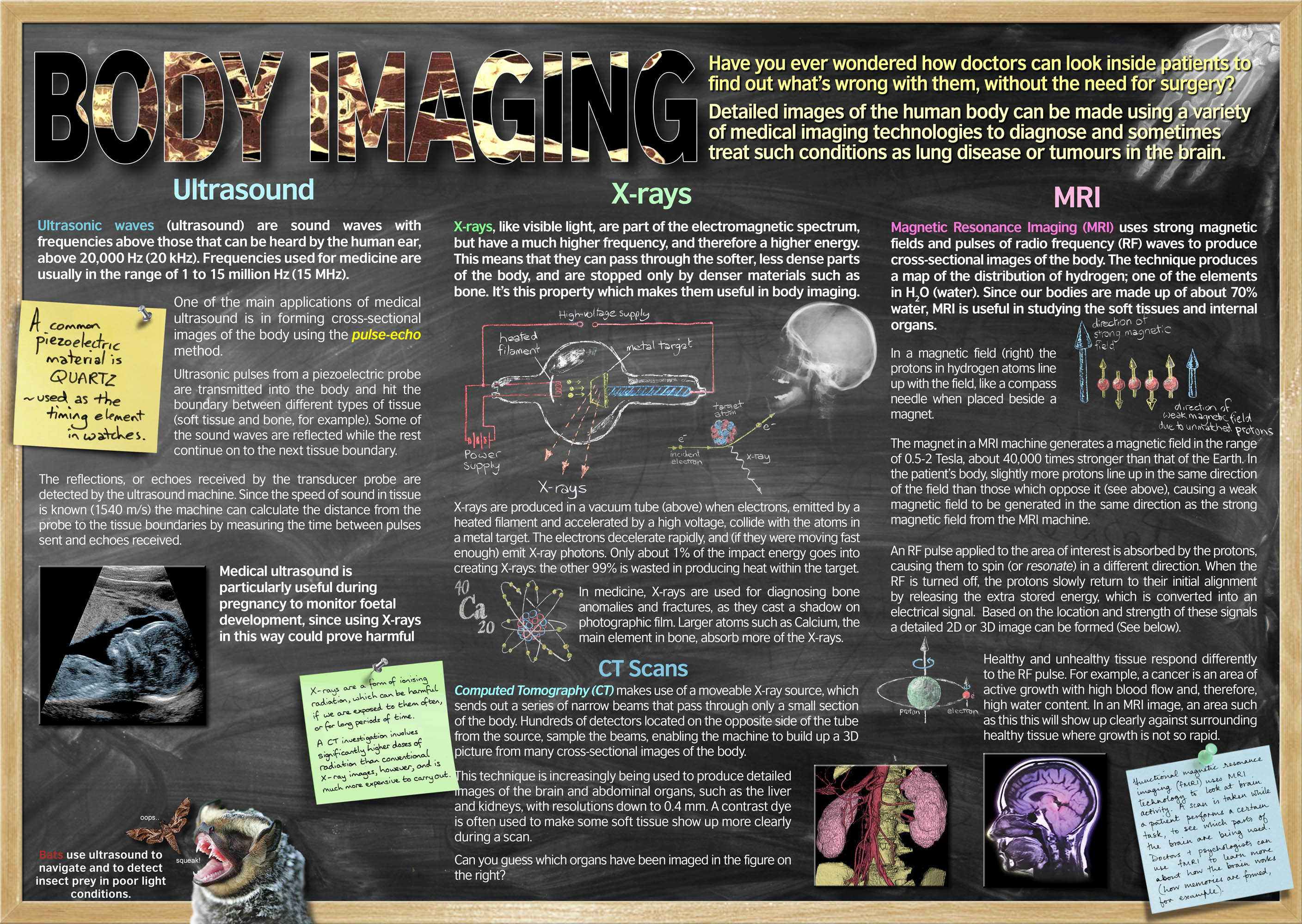
There are various methods of body imaging, and these are used for different tasks. Some of the main ones are ultrasound, X-rays and MRI.
X-rays are a form of radiation, like light and radiowaves. X-rays can pass through soft tissue, such as skin and muscle, but cannot pass through bone. This means that if we pass X-rays through a person onto photographic film, the bones will show up on the film. We can then see any breaks or damage to the bone.
If we take lots of X-ray images of an area, all at different angles, we can combine these images together using a computer to give a 3D model of the area. This is called CT scanning.
If you've ever had an X-ray, you'll know that the doctor or radiologist (a person who does X-rays) leaves the room for the X-ray. That is because X-rays can cause cancer. During your life, you will have so few X-rays that they will have less effect on you overall than sunlight. However radiologists will carry out many X-rays every day, so leave the room to limit their exposure.
Ultrasound uses sound to look inside our bodies. It is often used to look at babies in their mothers' wombs. A hand-held device emits really high-pitched sounds, so high that we can't hear them, on the woman's stomach. The sounds bounce off the baby and back to the device. The time taken for the sounds to bounce back allows a computer to construct an image of the baby. Ultrasound allows us to check that the baby is healthy and can determine if the baby is a girl or a boy.
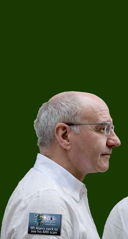
To look at soft tissue, such as our internal organs, we can use MRI scanning. This uses radiowaves and magnets to map the water in the body. This produces accurate and detailed images that can be very useful in diagnosis. The Body Imaging exhibit uses two MRI scans that were taken of a (now retired) senior lecturer in Physics, Alan Walker. If you roll the mouse over his head, below, you can see the lift-up flap that shows the MRI scan. Click on the scan images to see more.
Roll the mouse over the images above, and click on the scans to see more
If we are not able to diagnose illness by any of these methods, doctors may have to carry out exploratory surgery. That means they carry out an operation to have a look for signs of disease.
|
Questions
|
| 1 |
Body imaging is a way that doctors can ________ disease. |
| 2 |
What can't X-rays pass through in our bodies? |
| 3 |
What does a radiologist do? |
| 4 |
What can we find out about a baby from ultrasound? |
| 5 |
What substance in our bodies does MRI scanning map? |
| 6 |
Why do you think doctors want to avoid exploratory surgery? |
|
Activities
|
| 1 |
Sometimes you get to keep copies of X-rays you've had. If you've got some at home, have a look, otherwise try to find some on the internet. Have a look at some before and after shots, such as a broken bone then the same bone healed, or teeth before and after orthodontic treatment. Are the differences easy to see? |
| 2 |
Functional MRI scanning involves scanning people's brains whilst they carry out a particular task to see what happens in their brains. Try to think of some interesting tasks that people could do whilst having an MRI scan. Remember that scanners are not portable and there isn't much space in them. |
|